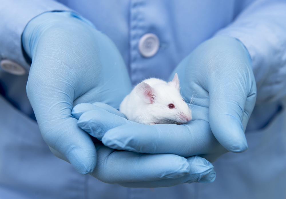
Sanfilippo Type A Changes Metabolism of Sugar and Fat Molecules in Brain Regions, Mouse Study Shows

Sanfilippo syndrome type A leads to changes in the metabolism of gangliosides (sugar-fat molecules) and sphingolipids (fat molecules) in different brain regions, and at different stages of the disease, according to a mouse study.
These findings pinpoint sphingolipids as potential therapeutic targets in people with this condition.
The study, “Sphingolipid dyshomeostasis in the brain of the mouse model of mucopolysaccharidosis type IIIA,” was published in the journal Molecular Genetics and Metabolism.
Sanfilippo syndrome type A, also called mucopolysaccharidosis type IIIA (MPS IIIA), is caused by a deficiency in the enzyme heparan N-sulfatase, which is responsible for the breakdown of a complex sugar molecule called heparan sulfate.
This condition leads to the accumulation of heparan sulfate inside lysosomes (the cell compartments responsible for digesting and recycling substances) inducing cell damage, mainly in the central nervous system (the brain and spinal cord).
In addition to heparan sulfate, other molecules are known to build up in these patients’ brains, namely GM2 and GM3 gangliosides, sugar-fat molecules derived from specific molecules of the sphingolipids family, a type of fat.
Gangliosides are most abundant in the brain, where they are involved in important processes related to nerve cells, and their accumulation has been hypothesized to cause changes in nerve cell structure, communication, and survival.
However, the specific regions of the brain where gangliosides accumulate, and whether this accumulation changes with disease progression, remain unclear.
Researchers set out to comprehensively analyze changes in ganglioside metabolism within the brain of the naturally occurring MPS IIIA mouse model at early and late stages of disease progression.
They also evaluated potential associations between the levels of gangliosides and heparan sulfate, and whether ganglioside-related sphingolipids were changed in this model.
The team analyzed four regions of the brain — the brain stem, cerebellum, cortex, and sub-cortex — in MPS IIIA mice at early (one month of age) and late (six months) stages of disease, and in age-matched healthy mice.
MPS IIIA mice showed high levels of GM2 and GM3 gangliosides in the brain stem, cerebellum, and sub-cortex, but not in the cortex, at early stage of disease, compared to healthy mice.
At late stage of disease, all brain regions showed significant buildup of GM2 and GM3 (particularly the brain stem), as well of another type of ganglioside, GD2, when compared to healthy mice.
“These gangliosides should not be expressed in the mature brain but it is still not clear what role, if any, they play in neurological demise,” the researchers wrote.
Mice at an early stage of disease showed a significant accumulation of heparan sulfate across all brain regions, which became more pronounced in the cortex and subcortex at a later stage.
Further analysis revealed several associations between the accumulation of these gangliosides and the levels of heparan sulfate in the distinct regions of the brain.
Results also showed that significant changes in sphingolipids levels were found mainly at later stages of disease in these mice.
High levels of ceramine and dihexosylceramide (DHC) — ganglioside precursors — were present mainly in the cortex and sub-cortex, while low levels of sphingomyelin, sulfatide, and DHC (all containing a specific structure called 24:1 fatty acid) were found in the brain stem.
Changes in DHC and ceramine levels have been associated with nerve cell dysfunction in several neurodegenerative diseases, and sphingolipids with 24:1 fatty acid are known to be involved in myelination — the formation of the protective layer around nerve cells that allows fast electric signal transmission.
The team noted that the occurrence of similar accumulation of gangliosides, DHC, and ceramine in the cortex and sub-cortex regions only at later stages of disease, suggests a potential association between changes in gangliosides’ and sphingolipids’ metabolisms and disease progression.
They also hypothesized that this ganglioside buildup may be due to an increase in its production, given the increased levels of DHC, its immediate precursor molecule.
Future studies are required to clarify the role of these metabolic changes in neurodegeneration and investigate sphingolipids as new potential targets in the treatment of Sanfilippo syndrome type A.


